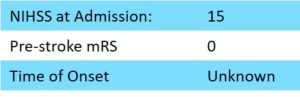
88 year old female patient presented with a NIHSS of 15.
The patient was found on the floor of her bathroom, with left side hemiplegia. Her pre-stroke mRS was assessed to be 0.
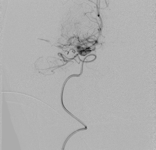
Native CT showed hyperdense M1 segment on the right side. CT angio confirmed M1 occlusion.
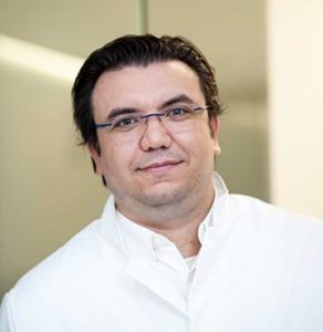
Dr. Vladimir Kalousek
Sisters Charity Hospital
Zagreb, Croatia
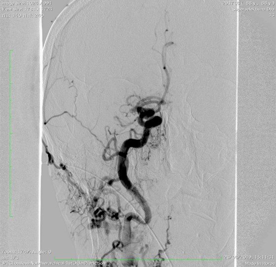
Dr Kalousek and the Sisters’ Charity team decided to treat the patient with thrombectomy using a NeVa M1 (4 mm x 30 mm) under distal aspiration.
The access set up was: 9F femoral sheath , 9F Cello , 0.064” aspiration catheter and 0.027” microcatheter.
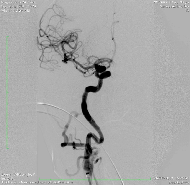
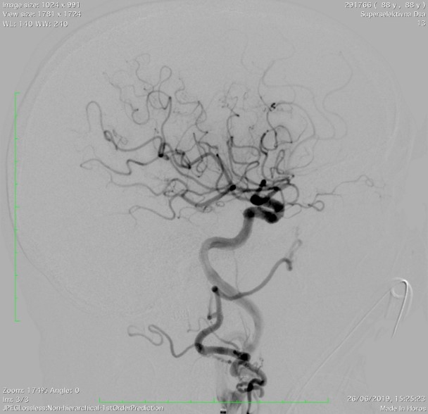
Full recanalization was possible in a single pass.
Thrombectomy was done under distal aspiration with NeVa M1 (4mm x 30mm). Distal aspiration was used as flow control strategy.
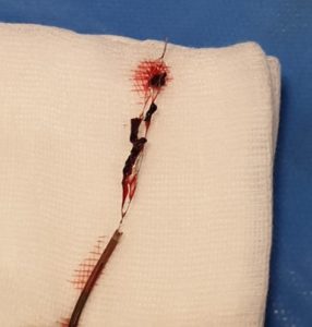
Full recanalization (TICI 3) was possible with a single pass and significant thrombus was observed throughout the body of NeVa and inside the distal tip.
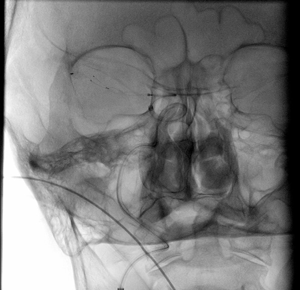
Control CT showed demarcation of the core of infarct in basal ganglia.
Post-procedure NIHSS of the patient was 6 and she began to recover immediately.
At VESALIO, we feel blessed to be part of the stroke field, where together with these dedicated physicians, we can make an incredible impact on people’s lives.
Thank you Zagreb !
| Cookie | Duration | Description |
|---|---|---|
| cookielawinfo-checkbox-analytics | 11 months | This cookie is set by GDPR Cookie Consent plugin. The cookie is used to store the user consent for the cookies in the category "Analytics". |
| cookielawinfo-checkbox-functional | 11 months | The cookie is set by GDPR cookie consent to record the user consent for the cookies in the category "Functional". |
| cookielawinfo-checkbox-necessary | 11 months | This cookie is set by GDPR Cookie Consent plugin. The cookies is used to store the user consent for the cookies in the category "Necessary". |
| cookielawinfo-checkbox-others | 11 months | This cookie is set by GDPR Cookie Consent plugin. The cookie is used to store the user consent for the cookies in the category "Other. |
| cookielawinfo-checkbox-performance | 11 months | This cookie is set by GDPR Cookie Consent plugin. The cookie is used to store the user consent for the cookies in the category "Performance". |
| viewed_cookie_policy | 11 months | The cookie is set by the GDPR Cookie Consent plugin and is used to store whether or not user has consented to the use of cookies. It does not store any personal data. |
by removing vascular occlusions and restoring blood flow
Please select your region to enter our world of innovation:
"*" indicates required fields