ONE-PASS RESCUE IN A CAROTID OCCLUSION INVOLVING P-COM & AChA
NeVa™ 5.5 x 37 mm
Dr. med. Christian Commodaro
Medico Capoclinica, INR Unit, Lugano, Switzerland
NeVa™ 5.5 x 37 mm
Dr. med. Christian Commodaro
Medico Capoclinica, INR Unit, Lugano, Switzerland
A 55-year-old male patient with a mechanical heart valve presented with a fluctuating NIHSS ranging from 3 to 11.

Dr. med. Christian Commodaro
Medico Capoclinica, INR Unit
Lugano, Switzerland
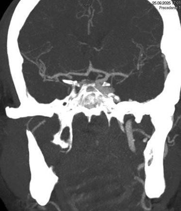
1. Occlusion of the right internal carotid artery (R-ICA intracranial segment) involving P-Com & AChA.
2. Occlusion of the right P-Com in the context of a fetal type PCA.
3. A patent apex of the right carotid artery with normal intracranial circulation elsewhere.


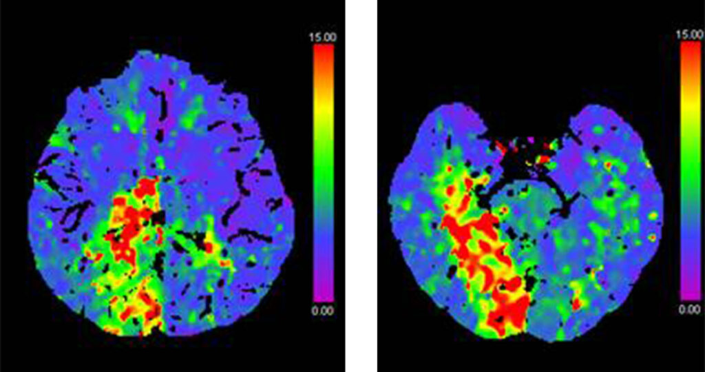
CT Perfusion maps (T-MAX) showing hypo-perfused posterior cerebral artery and AChA territories on the right side.
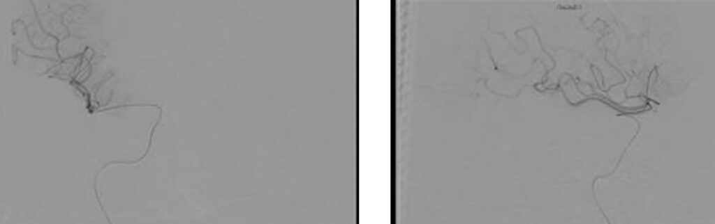
Angiographic AP and LL views showing blood flow arrest in the R-ICA after the bulb (“stump flow”) due to the occlusion of the intracranial segment of the ICA

AP and LL images from the roadmap during the navigation across the occluded segment.

Selective injection into the right M1 segment to confirm the correct distal position of the microcatheter.
1st Pass:
Direct aspiration with a .071″ aspiration catheter
2nd Pass:
A different stent retriever (5 x 37 mm) associated with distal aspiration with the .071″ aspiration catheter (SAVE technique)
3rd and final pass: with NeVa 5.5 x 37mm associated with distal aspiration with the .071″ aspiration catheter (SAVE technique):
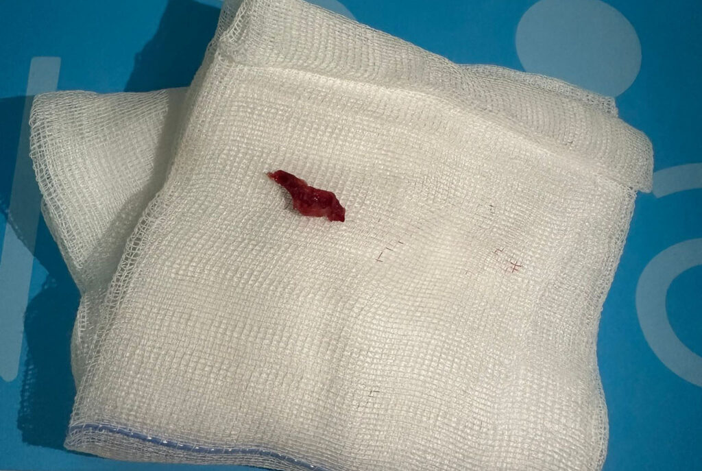
Two unsuccessful thrombectomy attempts were made prior to NeVa use. The NeVa pass achieved complete recanalization with no distal thromboembolism (TICI 3):
1st pass success:
TICI 3 in a single pass with NeVa 5.5 x 37mm

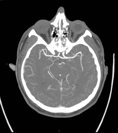
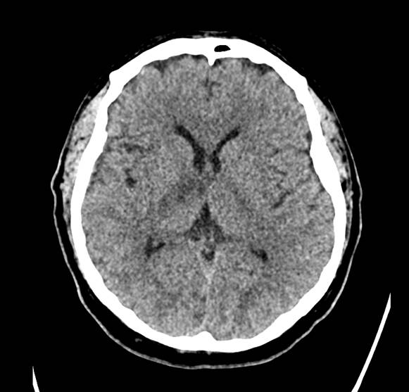
24-hr CT showed a small lesion in the anterior portion of the right thalamus and a marginal lesion in the adjacent right internal capsule
| Cookie | Duration | Description |
|---|---|---|
| cookielawinfo-checkbox-analytics | 11 months | This cookie is set by GDPR Cookie Consent plugin. The cookie is used to store the user consent for the cookies in the category "Analytics". |
| cookielawinfo-checkbox-functional | 11 months | The cookie is set by GDPR cookie consent to record the user consent for the cookies in the category "Functional". |
| cookielawinfo-checkbox-necessary | 11 months | This cookie is set by GDPR Cookie Consent plugin. The cookies is used to store the user consent for the cookies in the category "Necessary". |
| cookielawinfo-checkbox-others | 11 months | This cookie is set by GDPR Cookie Consent plugin. The cookie is used to store the user consent for the cookies in the category "Other. |
| cookielawinfo-checkbox-performance | 11 months | This cookie is set by GDPR Cookie Consent plugin. The cookie is used to store the user consent for the cookies in the category "Performance". |
| viewed_cookie_policy | 11 months | The cookie is set by the GDPR Cookie Consent plugin and is used to store whether or not user has consented to the use of cookies. It does not store any personal data. |
by removing vascular occlusions and restoring blood flow
Please select your region to enter our world of innovation:
"*" indicates required fields