NeVa Saves the Day after 5 Failed Passes
NeVa™ 4.5 x 37 mm
Bucharest University Emergency Hospital Stroke Team
NeVa™ 4.5 x 37 mm
Bucharest University Emergency Hospital Stroke Team
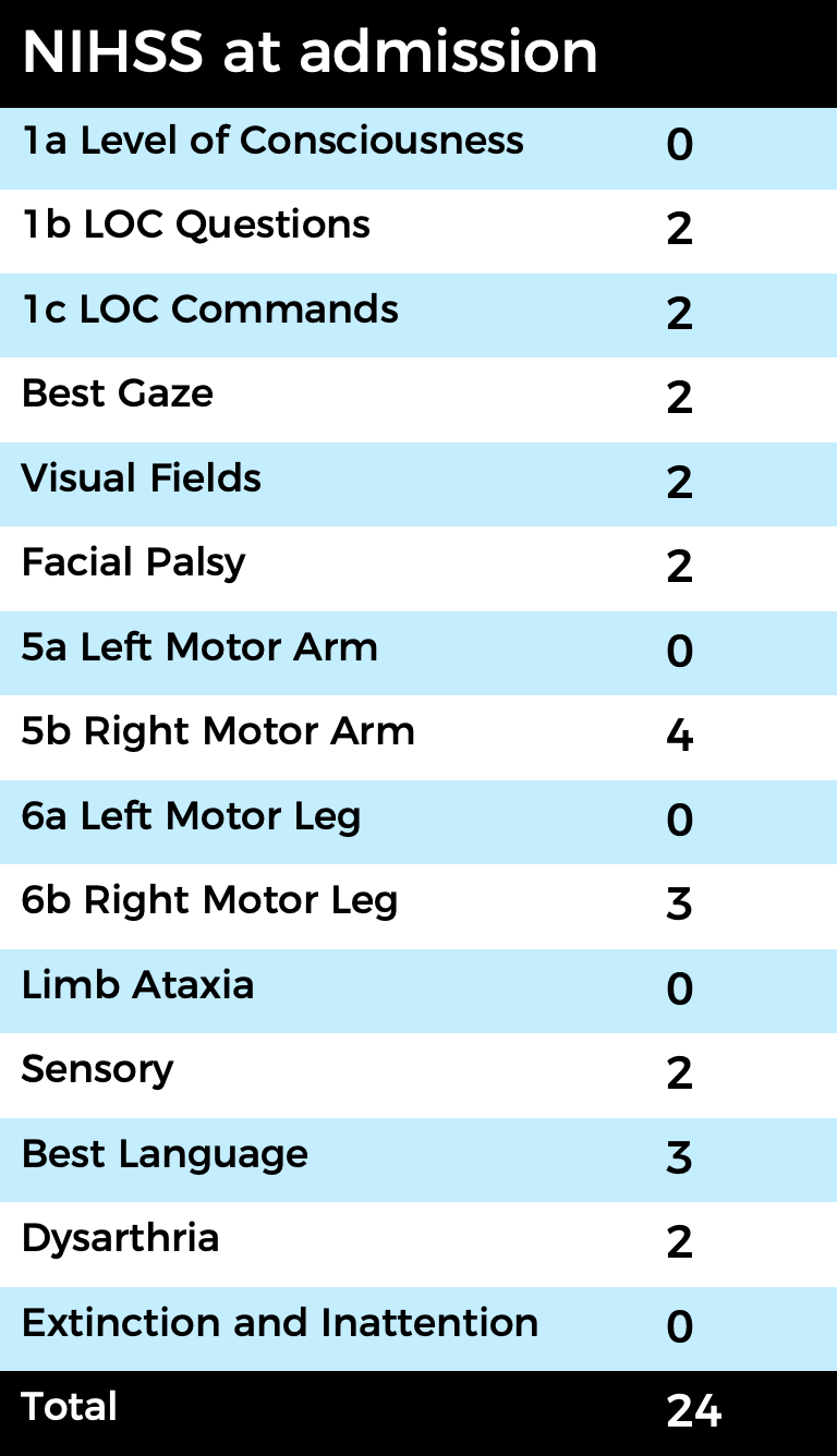
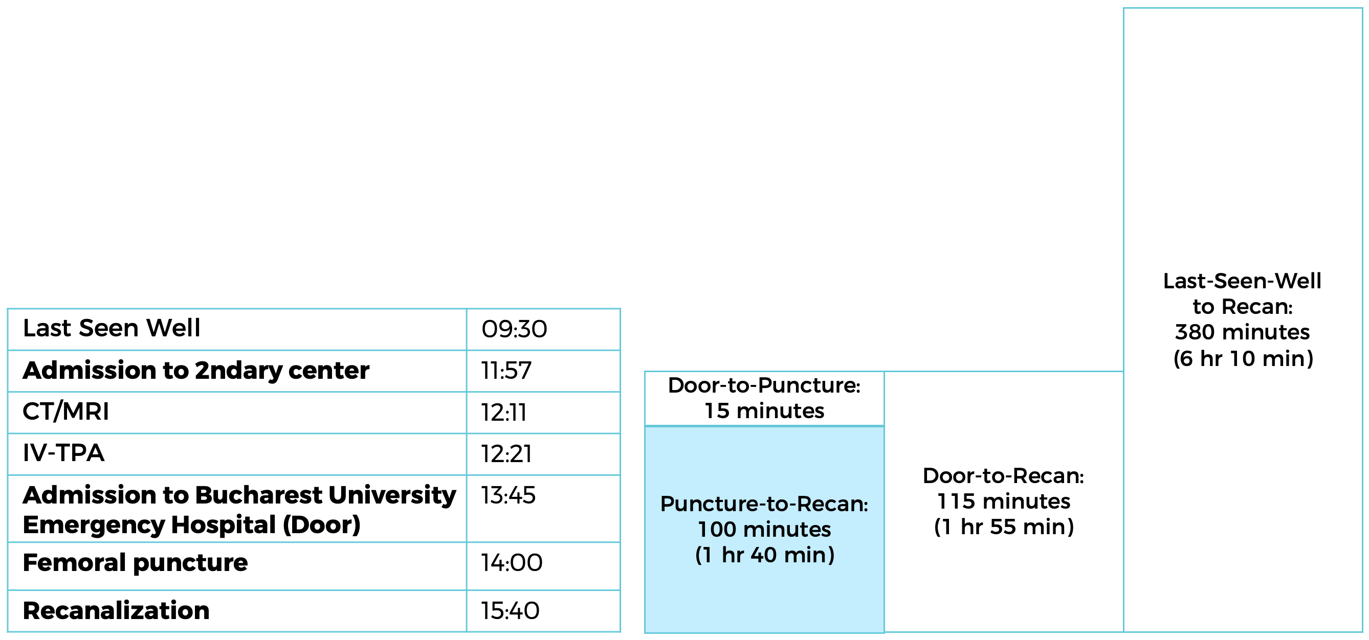
80 year old male patient presented with a NIHSS of 24, 4 hr 15 min after symptom onset.
The patient was transferred to the Bucharest University Emergency Hospital Stroke Unit after tPA administration in secondary center.
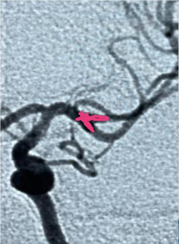
Imaging confirmed occlusion of the left M1 segment.
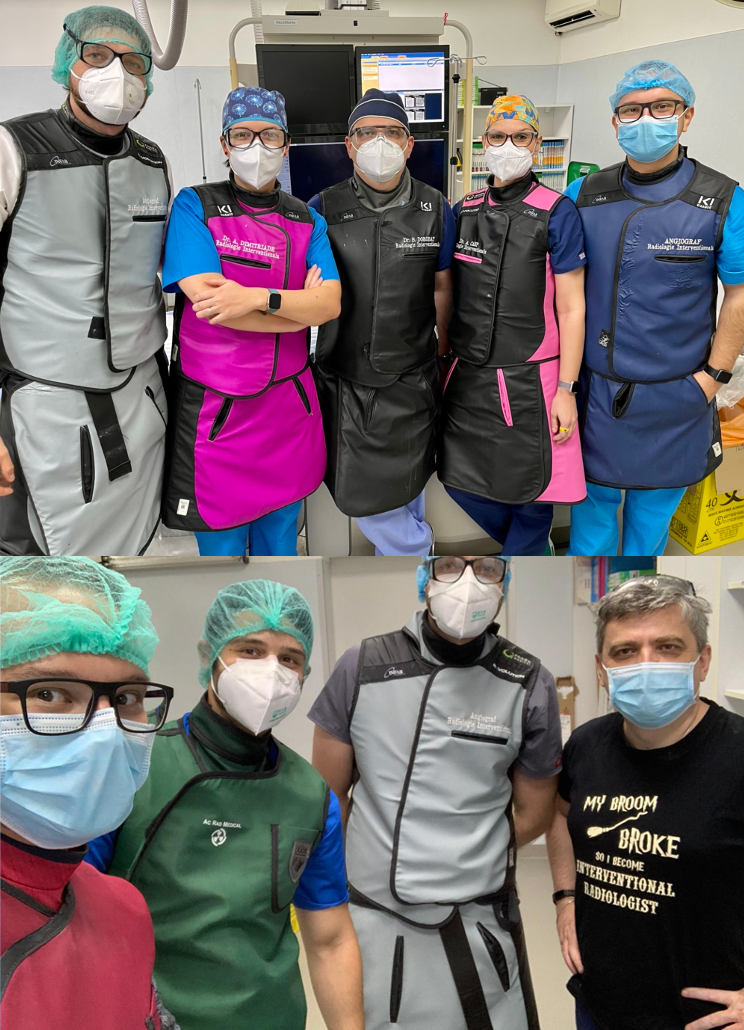
Dr. Bogdan DOROBĂȚ,
Dr. Adela DIMITRIADE, Dr. Alexandra CARP, Dr. Andrei SIMONOV, Dr. Mihai IONESCU
Bucharest University Emergency Hospital
Bucharest/Romania
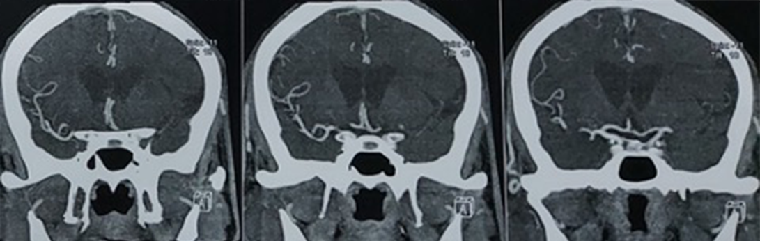
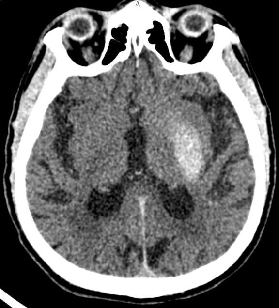
Pre-op CT confirmed acute ischemic stroke with some early ischemic changes in the deep left MCA territory. ASPECTS score of 7
IV-tPA was administered at secondary center prior to the patient being transferred to the Bucharest University Emergency Hospital Stroke for mechanical thrombectomy.
Femoral approach was used for access and the team decided to use a 9-French balloon guide catheter (BGC) as a flow control strategy. Aspiration was done through the BGC during retrieval. A 0.027” microcatheter was used for device delivery.
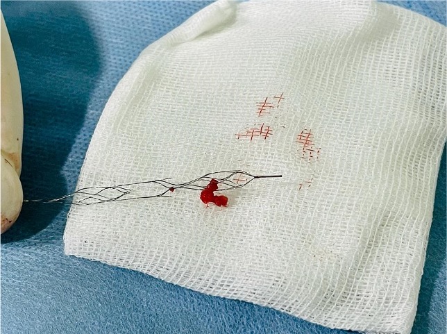
5 thrombectomy passes were done with no improvement on the initial TICI-0 occlusion status:
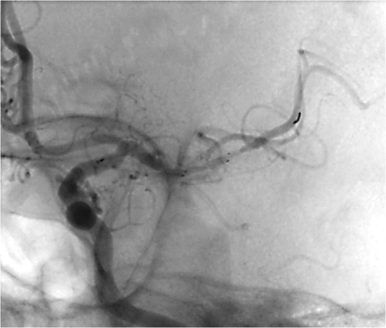
For the 6th pass, a Neva 4.5 x 37 mm was used. Initially, NeVa was incorrectly positioned (too proximally – thrombus at level of distal basket). After correct positioning, complete recanalization (TICI 3) of the occlusion was achieved with a single pass.
9F Balloon Guide Catheter was placed in distal ICA. After 5 successive passes with two different stent-retrievers (4.0 x 32 mm and 6.0 x 40 mm) the initial left-M1 occlusion remained in place.
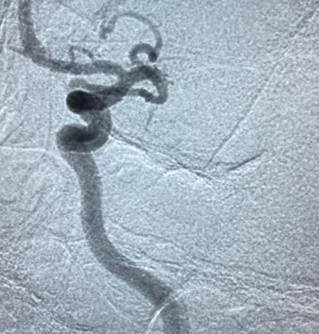
Initial angio showing the left-M1 occlusion
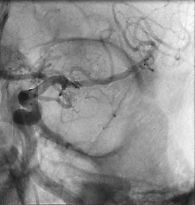
A pass with a 4.0 x 32 mm stent retriever positioned in the MCA
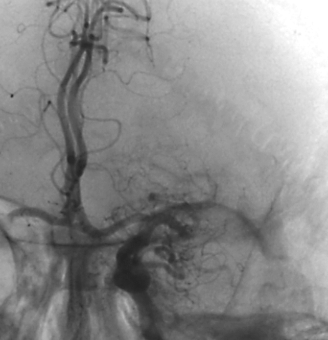
NeVa 4.5 x 37 mm positioned across the upper branch of the MCA

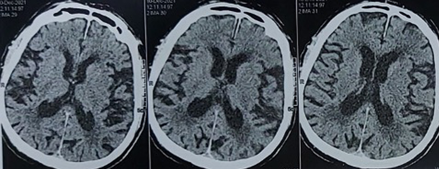
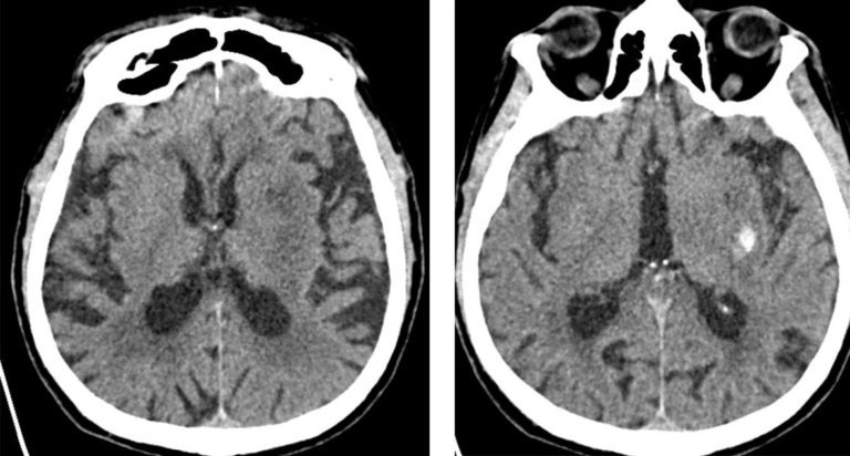
12-hour Post-op CT shows contrast staining of the lenticular nucleus
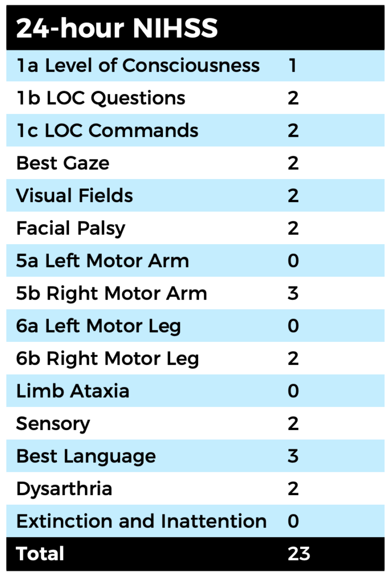
24 hour NIHSS of the patient was 23, which later improved to 18.
The patient remained hospitalized with signs of aspiration pneumonia unrelated to the thrombectomy.
At VESALIO, we feel blessed to be part of the stroke field where together with these dedicated stroke teams, we can make an incredible impact on people’s lives.
Thank you Bucharest University Emergency Hospital stroke team: Dr. Bogdan DOROBĂȚ, Dr. Adela DIMITRIADE, Dr. Alexandra CARP, Dr. Andrei SIMONOV, and Dr. Mihai IONESCU.
| Cookie | Duration | Description |
|---|---|---|
| cookielawinfo-checkbox-analytics | 11 months | This cookie is set by GDPR Cookie Consent plugin. The cookie is used to store the user consent for the cookies in the category "Analytics". |
| cookielawinfo-checkbox-functional | 11 months | The cookie is set by GDPR cookie consent to record the user consent for the cookies in the category "Functional". |
| cookielawinfo-checkbox-necessary | 11 months | This cookie is set by GDPR Cookie Consent plugin. The cookies is used to store the user consent for the cookies in the category "Necessary". |
| cookielawinfo-checkbox-others | 11 months | This cookie is set by GDPR Cookie Consent plugin. The cookie is used to store the user consent for the cookies in the category "Other. |
| cookielawinfo-checkbox-performance | 11 months | This cookie is set by GDPR Cookie Consent plugin. The cookie is used to store the user consent for the cookies in the category "Performance". |
| viewed_cookie_policy | 11 months | The cookie is set by the GDPR Cookie Consent plugin and is used to store whether or not user has consented to the use of cookies. It does not store any personal data. |
by removing vascular occlusions and restoring blood flow
Please select your region to enter our world of innovation:
"*" indicates required fields