86-year-old female patient presenting with a NIHSS of 12 was referred with an IV-tPA (drip & ship) from the primary care centre.
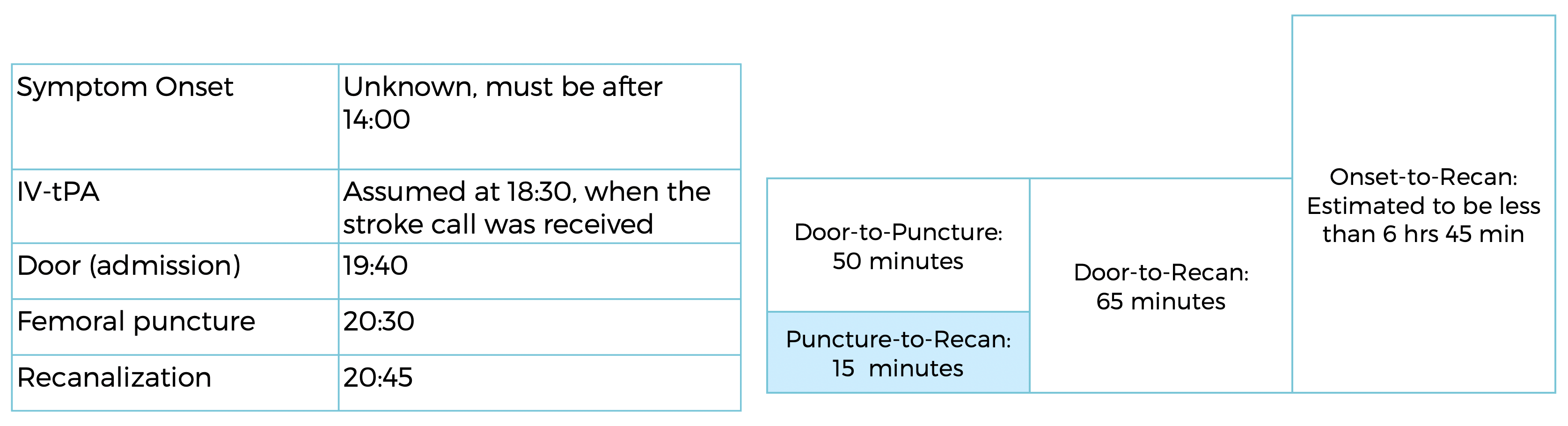
The exact time of symptom onset is unknown, but estimated to be after 14:00.
The stroke alert was received at 18:30.
At 19:40, when the patient got admitted to the Sisters Charity Stroke Unit, her neurological status had further deteriorated.
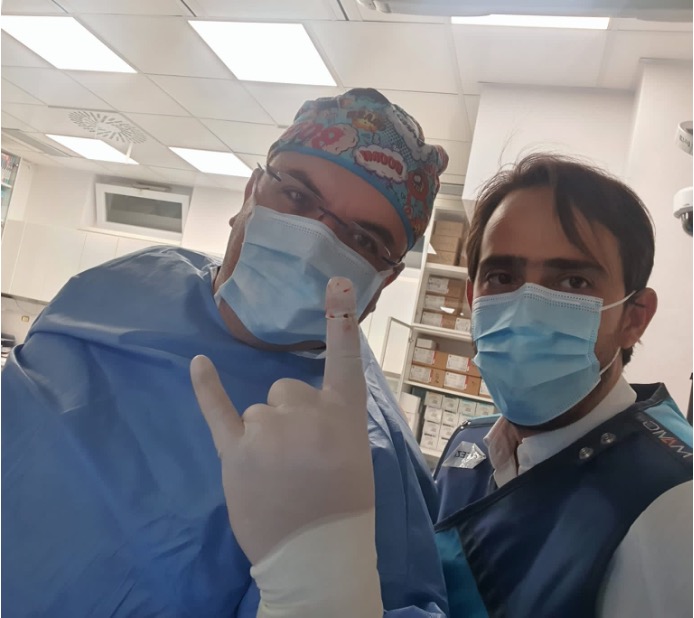
Dr. Dr Vladimir KALOUSEK UHC Sisters of Charity, Zagreb, CROATIA
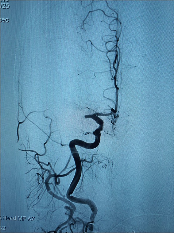
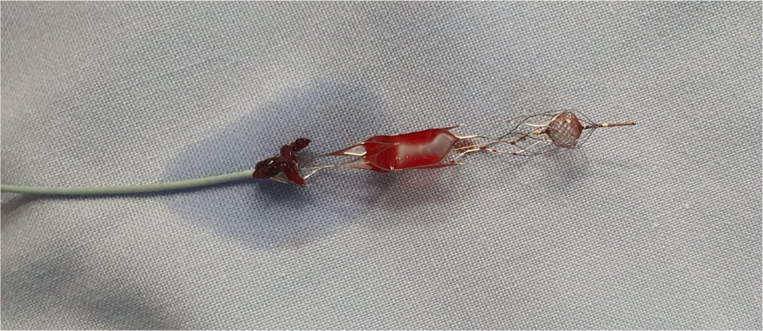
Dr Kalousek and team decided to proceed to thrombectomy, which was done under distal aspiration with a NeVa NET 5.5 x 37 mm
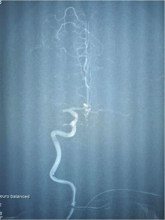
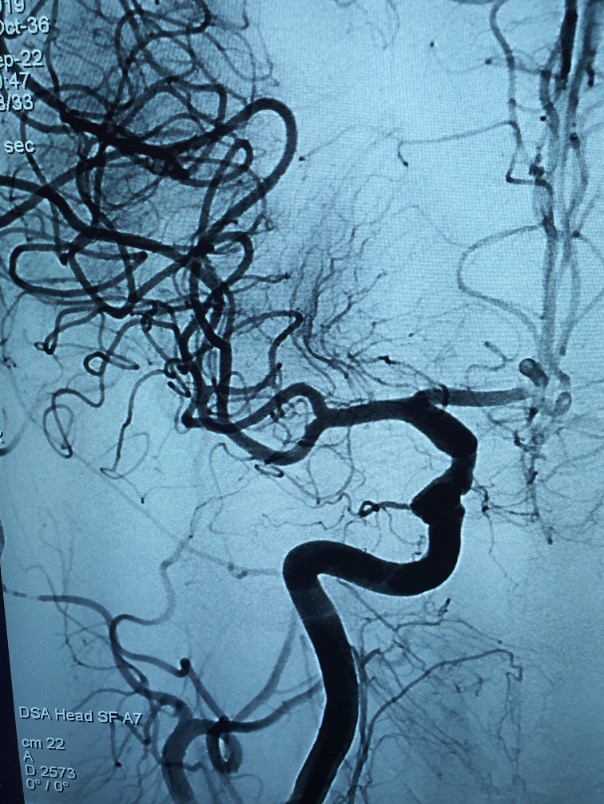
ANGIO SHOWING THE Right-M1 OCCLUSION
Angiographic imaging at the beginning of the case confirmed the occlussion of the right-M1 branch.
NeVa NET 5.5 x 37 mm was deployed across
the M1 SEGMENT
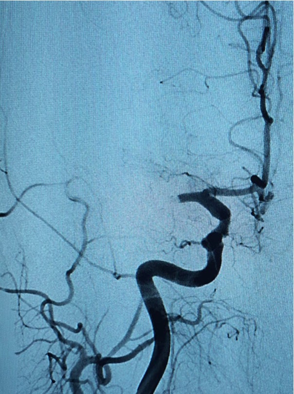
NeVa NET 5.5 x 37 mm was taken partially into the
DAC during retrieval
BEFORE
AFTER
Thrombectomy of the Right-M1 lesion was done using a NeVa NET 5.5 x 37 mm, delivered within a 0.027” micro-catheter under co-aspiration with a 0.071” ID-DAC.
Full recanalization (TICI 3) was achieved in the first pass and thrombus was observed within the device.
The patient was discharged with an NIHSS of 3.
At VESALIO, we feel blessed to be part of the stroke field where together with these dedicated stroke teams, we can make an incredible impact on people’s lives.
Thank you Zagreb sister’s Charity stroke team: Dr Kalousek, all the lab technicians and nurses as well as the anesthesiology team supporting this case.
| Cookie | Duration | Description |
|---|---|---|
| cookielawinfo-checkbox-analytics | 11 months | This cookie is set by GDPR Cookie Consent plugin. The cookie is used to store the user consent for the cookies in the category "Analytics". |
| cookielawinfo-checkbox-functional | 11 months | The cookie is set by GDPR cookie consent to record the user consent for the cookies in the category "Functional". |
| cookielawinfo-checkbox-necessary | 11 months | This cookie is set by GDPR Cookie Consent plugin. The cookies is used to store the user consent for the cookies in the category "Necessary". |
| cookielawinfo-checkbox-others | 11 months | This cookie is set by GDPR Cookie Consent plugin. The cookie is used to store the user consent for the cookies in the category "Other. |
| cookielawinfo-checkbox-performance | 11 months | This cookie is set by GDPR Cookie Consent plugin. The cookie is used to store the user consent for the cookies in the category "Performance". |
| viewed_cookie_policy | 11 months | The cookie is set by the GDPR Cookie Consent plugin and is used to store whether or not user has consented to the use of cookies. It does not store any personal data. |
by removing vascular occlusions and restoring blood flow
Please select your region to enter our world of innovation:
"*" indicates required fields